Schematic depiction of the distribution of the PV autoantigens Dsg1

Download scientific diagram | | Schematic depiction of the distribution of the PV autoantigens Dsg1 (green) and Dsg3 (red) and the composition of desmosome along different epidermal layers in normal epidermis (left) and PV-affected epidermis (right). *Significant difference to the value which is indicated that it is compared to. from publication: Dsg1 and Dsg3 Composition of Desmosomes Across Human Epidermis and Alterations in Pemphigus Vulgaris Patient Skin | Desmosomes are important epidermal adhesion units and signalling hubs, which play an important role in pemphigus pathogenesis. Different expression patterns of the pemphigus autoantigens desmoglein (Dsg)1 and Dsg3 across different epidermal layers have been demonstrated. | Desmosomes, Pemphigus and Epidermis | ResearchGate, the professional network for scientists.

The Immune System in Normal Skin and in AIBD

Desmoglein-Specific B-Cell−Targeted Single-Cell Analysis Revealing Unique Gene Regulation in Patients with Pemphigus - ScienceDirect

A) Schematic depiction of a desmosome unit sub-cell with all
Life, Free Full-Text
Desmosome assembly, homeostasis, and desmosomal disease - Document - Gale Academic OneFile
caba-10k_20211231.htm

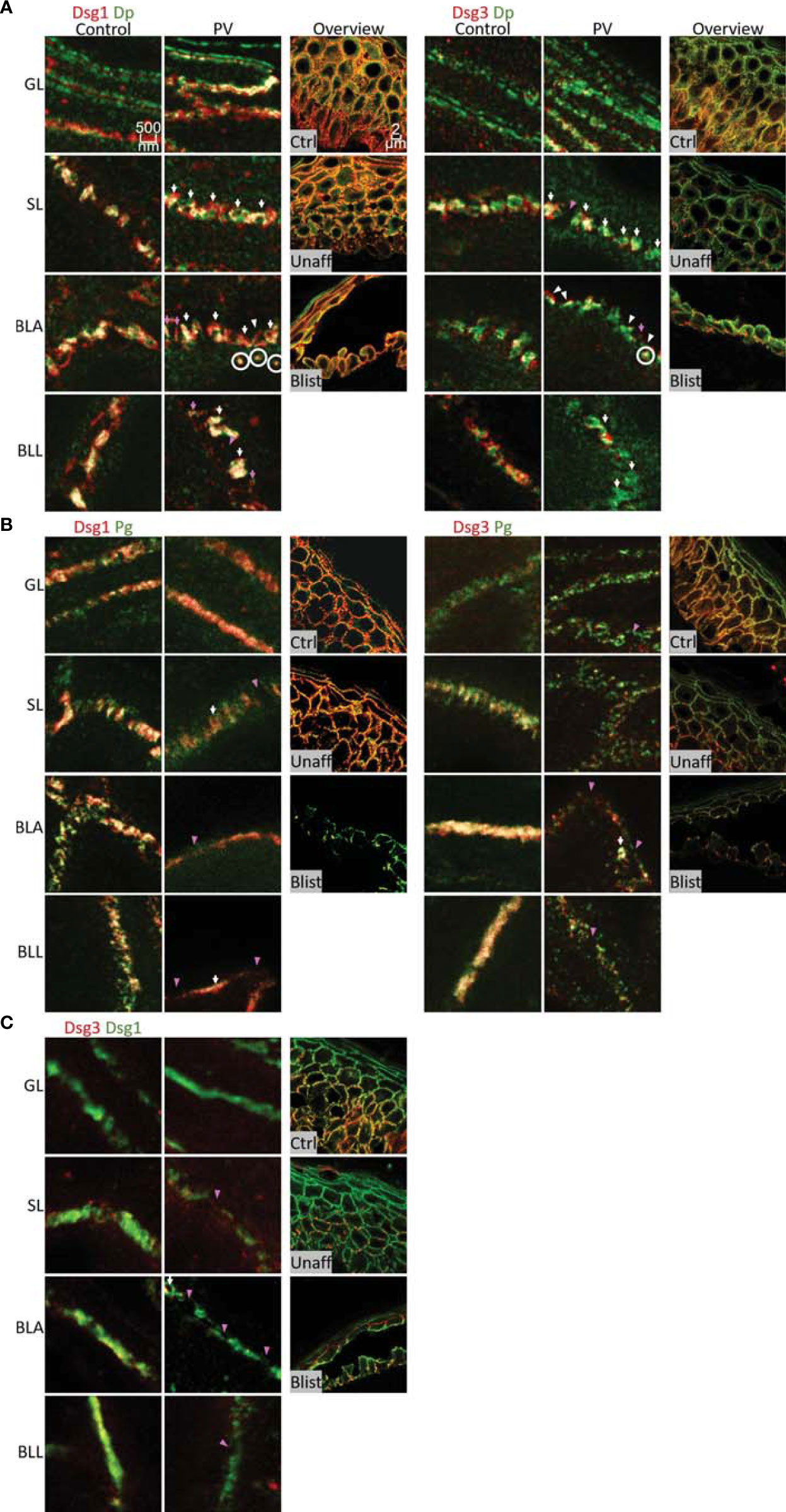
Different signaling patterns contribute to loss of keratinocyte

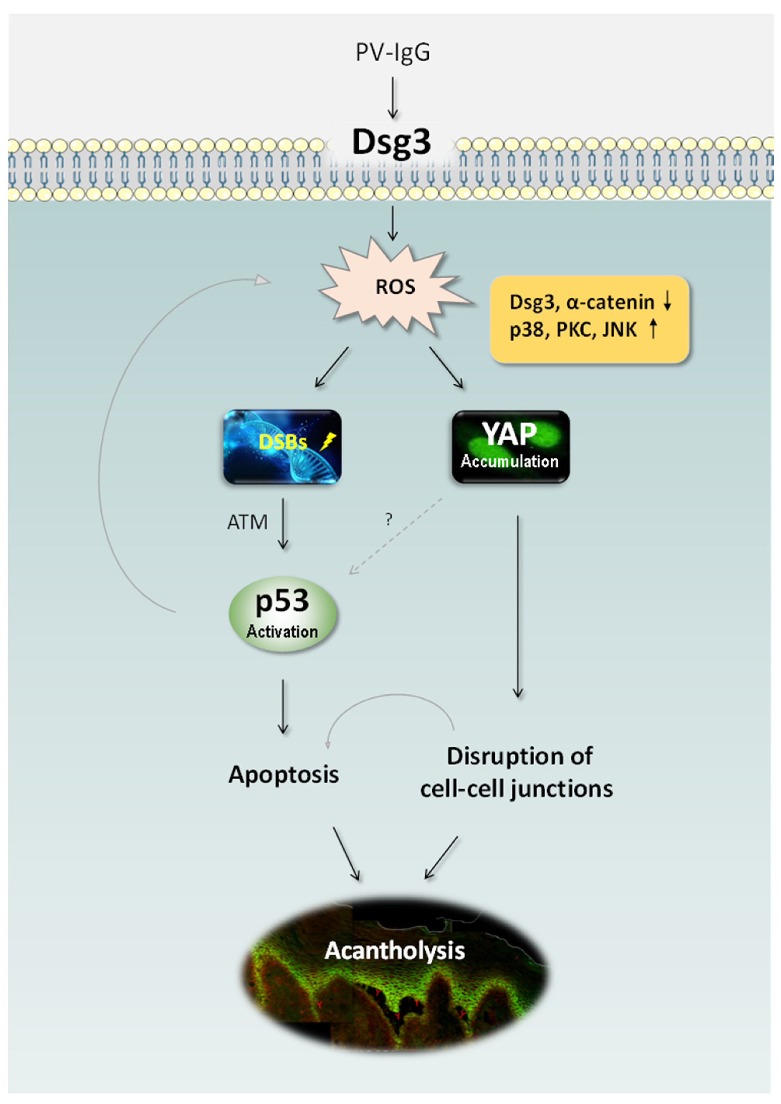
Up-regulation of ST18 in pemphigus vulgaris drives a self-amplifying p53-dependent pathomechanism resulting in decreased desmoglein 3 expression
Frontiers Dsg1 and Dsg3 Composition of Desmosomes Across Human Epidermis and Alterations in Pemphigus Vulgaris Patient Skin

Jens WASCHKE, Ludwig-Maximilians-University of Munich, München, LMU, Institute for Anatomy and Cell Biology

Epistasis between DSG1 and HLA class II genes in pemphigus
A New Solid-Phase Immunosorbent for Selective Binding of Desmoglein 3 Autoantibodies in Patients with Pemphigus Vulgaris - Abramova - Acta Naturae
Cells, Free Full-Text

How does acantholysis occur in pemphigus vulgaris: a critical review - Lanza - 2006 - Journal of Cutaneous Pathology - Wiley Online Library