Optical microscope images, (a) 0h, (b) 16h, (c) 26h, (d) 38h and (e) 48h


A “D-π-A” type NiIIporphyrin-BODIPY hybrid: Expansion of visible-light absorption and antitumor activity of lymphoma cells - ScienceDirect

PDF) Homogenization heat treatment influence on microstructure

Optical microscope images, (a) 0h, (b) 16h, (c) 26h, (d) 38h and (e) 48h

Optical microscope images, (a) 0h, (b) 16h, (c) 26h, (d) 38h and (e) 48h

Optical microscope images: (a) screen-printed layout 1 (scale bar: 200

A) Schematic of in situ ZIF-8 growth on PLA fibers and Scanning
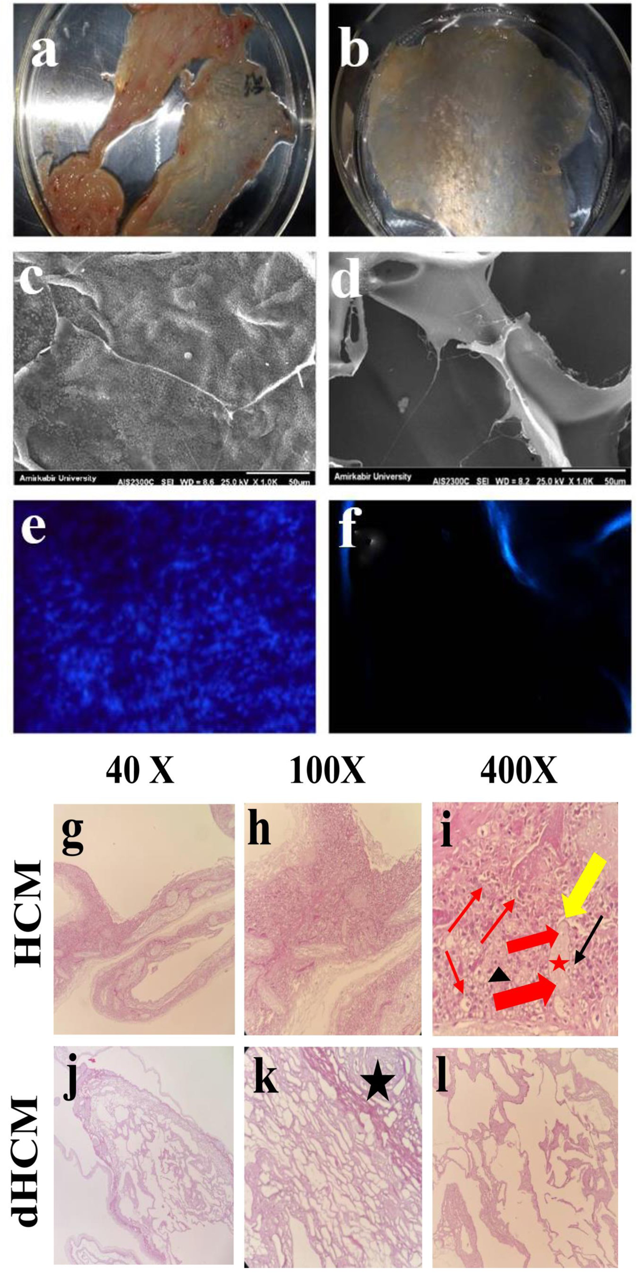
Biological study of skin wound treated with Alginate/Carboxymethyl cellulose/chorion membrane, diopside nanoparticles, and Botox A
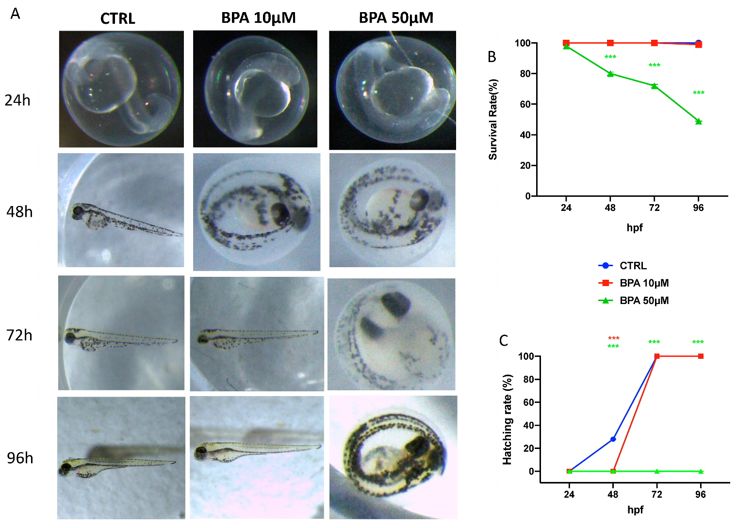
Toxics, Free Full-Text

Pharmaceuticals, Free Full-Text

Pharmaceuticals, Free Full-Text

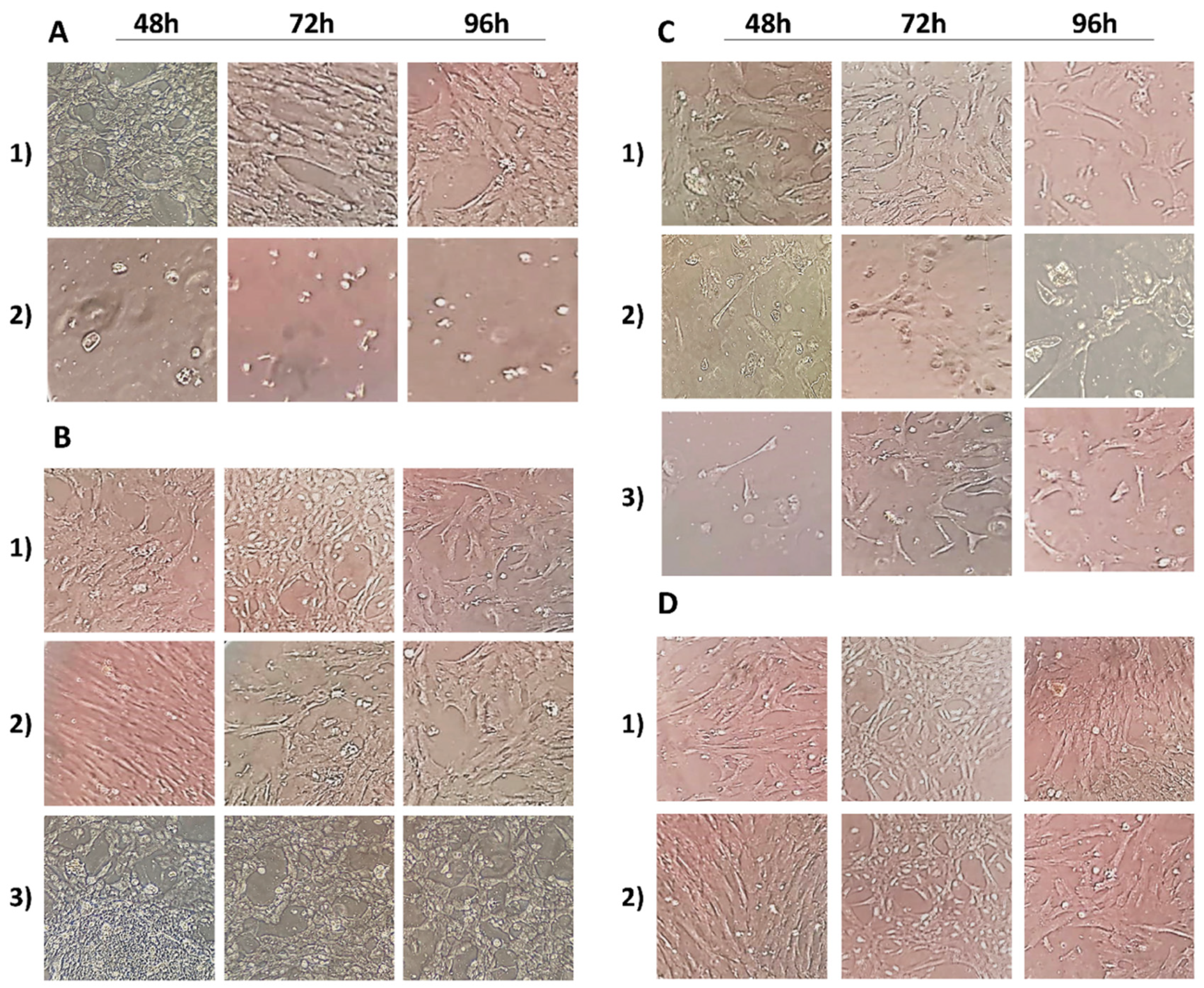
Optical images of the cell morphology using an optical microscope in
Antioxidants, Free Full-Text

Accelerated wound closure rate by hyaluronic acid release from coated PHBV electrospun fiber scaffolds - ScienceDirect

Light microscopy (20 × by 10 × ) of the cross-sections of mid small