Optical Coherence Tomography: Imaging Mouse Retinal Ganglion Cells In Vivo

Scientific Article | Structural changes in the retina are common manifestations of ophthalmic diseases.
Longitudinal In Vivo Imaging of Retinal Ganglion Cells and Retinal Thickness Changes Following Optic Nerve Injury in Mice
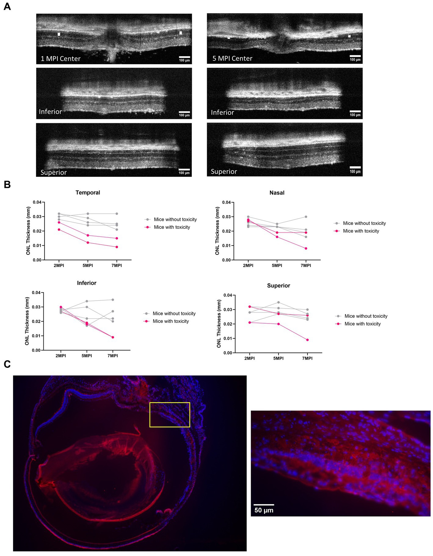
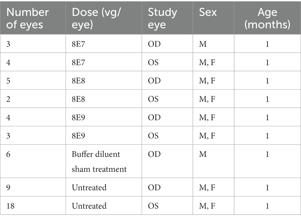
Frontiers The dose-response relationship of subretinal gene therapy with rAAV2tYF-CB-hRS1 in a mouse model of X-linked retinoschisis
Retina Tool - ImageJ-macros - MRI's Redmine

Longitudinal analysis of retinal ganglion cell damage at individual axon bundle level in mice using visible-light optical coherence tomography fibergraphy
Retinal Optical Coherence Tomography Imaging

Genes, Free Full-Text
Frontiers The dose-response relationship of subretinal gene therapy with rAAV2tYF-CB-hRS1 in a mouse model of X-linked retinoschisis

In vivo imaging of the inner retinal layer structure in mice after eye-opening using visible-light optical coherence tomography - ScienceDirect

Retinal Ganglion Cells: Methods and Protocols

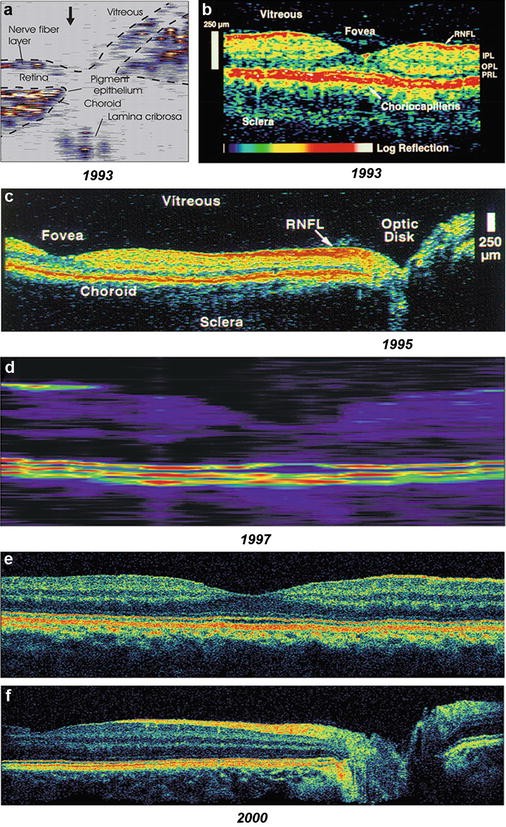
Fig. 9.10, [In vivo confocal reflectance and]. - High Resolution Imaging in Microscopy and Ophthalmology - NCBI Bookshelf